Chest findings in scleroderma

A 73 year old man with underlying scleroderma presenting with progressive shortness of breath.
A Chest X-Ray was performed.

  • Fibrotic changes involving the lower two thirds of the lung, with associated volume loss and honeycombing.
  • Ellevation of diaphragms suggestive of restrictive lung disease.
  • Subpleural reticular or reticulonodular opacity.
  • Cardiomegaly.

DISCUSSION:

Plain film:

  • insensitive to early changes
  • may be normal despite respiratory function test abnormalities.
  • pulmonary fibrosis 
  • dilated oesophagus
  • eggshell calcification of mediastinal nodes
  • pleural effusions are uncommon
  • enlargement of cardiac silhouette and pulmonary arteries due to scleroderma induced pulmonary vascular disease may also be evident. 

HRCT Thorax:

Azygous fissure


Clinical data: A foreign worker came for routine medical examination.

CHEST RADIOGRAPH (PA ERECT):


Findings:

  • Accessory fissure forming a thin ‘inverted comma’ line.
  • Azygous vein forming the head of a ‘tadpole’.

Disccusion:

    • an accessory fissure occurs in approximately 1-2% of individuals.
    • It is typically an incidental radiographic finding.
    • It has no associated signs or symptoms.
    • No treatment is necessary, as this is a normal anatomic variant.
    • vein appears to run within the lung wrapped by four layers of pleura (two parietal layers and two visceral layers) giving the appearance of ‘head of a tadpole’.
    • the azygous vein has a more lateral course and descends along a fissure in the right upper lobe, resulting in azygous fissure.
    • The azygos vein and trigone also are often seen.
      • Azygos vein: Appears as an ovoid, tear-shaped opacity in the inferior aspect of the accessory fissure.
      • Trigone: Appears as a triangular opacity that marks the superior aspect of azygos fissure.
    • accessory fissures of the lung usually occur at the borders of bronchopulmonary segments. They are normal variants.
    • other accessory fissures include inferior accessory fissure,  superior accessory fissure and left minor fissure.
    • the inferior accessory fissure surrounds the medial basal segment of the lower lobe.
    • the superior accessory fissure seperates the superior segment of lower lobe from basal segments.
    • left minor fissure separates the lingula from the rest of the left upper lobe. 
    • accessory fissures, like all other fissures, serve not only as natural barriers against infection but also help in localizing any focal pulmonary parenchymal disease.
  • Major fissures are oblique fissures and right transverse fissure.
  • The left lung is divided into two lobes, upper and lower. These lobes have their own pleural covering and these lie together to form the oblique (major) fissure.
  • The right lung, there is an oblique fissure and a horizontal fissure, separating the lung into three lobes – upper, middle, and lower. 
  • The horizontal fissure (right) is often seen on a normal frontal view CXR whereas the oblique fissures are often seen on a normal lateral view

CT THORAX:

Foreign bodies in the body

CASE 1

CLINICAL  DATA: Swallowing of coin.

  • Day 2 of coin swallowing.

  • Day 4 of coin swallowing.

CASE 2

CLINICAL DATA: Accidental injury. Wire penentrating the thigh.

  • A small piece of wire in the muscle of the thigh.

CASE 3

CLINICAL DATA: Coin swallowing.

  • 7 years old boy swallowed a 20 cent coin. Day 1

  • Day 2.

CASE 4

  • A worker with nail in his hand.

CASE 5

CLINICAL DATA:  A 47 year old lady came for routine Chest X-ray and Mammogram studies.History of insertion of ‘golden-threads’ insertion into the soft tissue of the body with the purpose of improving general well beings.

CASE 6

CLINICAL DATA: A one-year old boy swallowed an earring.

Abdominal radiograph showed:

CASE 7

CLINICAL DATA: A 37 year-old lady swallowed chicken bone while having lunch.

  • position of chicken bone at C6-C7 vertebral levels.
  • Impacted esophageal foreign body tends to occur at areas of esophageal luminal narrowing, either:

    a. physiologic/anatomic (i.e. gastroesophageal junction, aortic arch, left main bronchus, & cricopharyngeus muscle [C6 level]) or

    b. pathologic (i.e. stricture, tumor, Schatzki ring, & esophageal webs).

    More frequent in children than adults (80% vs. 20%, respectively).

    Removal is recommended within the first 24 hours, due to an increased risk of perforation with foreign bodies retained longer than 1 day.

    CT imaging may be helpful in select cases to delineate foreign bodies, evaluate for secondary signs like tissue emphysema, hematoma, or abscess, or to exclude subtle perforation.

    References:

    1. Federle MP, Guliani-Chabra S. Foreign body, esophagus. Statdx. Accessed June 30, 2010.

CASE 8

  • Position of the nail penentration.

Antiphospholipid syndrome (APS) with pulmonary embolism and DVT of lower limb.

CLINICAL DATA: 

A 29 years-old gentleman presented with sudden onset of chest pain, palpitation and reduced effort tolerance.

No abnormality of the lower limbs. CNS is unremarkable.

D-Dimers significantly elevated.

CHEST RADIOGRAPH (PA ERECT VIEW):


  • Enlarged pulmonary artery (15% of pulmonary embolism cases).
  • Fleischner sign – local widening of artery by clot/pulmonary hypertension.
  • Heart size, lung and pleural space are unremarkable.

CTA PULMONARY:


  • Thrombus in multiple segments of pulmonary arteries bilaterally (right main pulmonary artery, lobar and segmental arteries bilaterally).
  • No pleural effusion. No abnormality of the lungs.

ULTRASOUND VENOUS DOPPLER OF LOWER LIMB:

  • Acute hypoechoic thrombus in the left popliteal vein extending to distal 1/3 of left superficial femoral vein.
  • The affected segment of the vein is dilated and non compressible.

BLOOD TESTS:

  • VDRL positive.
  • Thrombocytopenia
  • Anticardiolipin Ig G Strongly +ve (118.1 GPL untis)
  • Anticardiolipin IgM antibody 4.7 MPL untis.

DIAGNOSIS:

Antiphospholipid syndrome with pulmonary embolism and DVT of left lower limb.

DISCUSSION:

  • a disorder that manifests clinically as recurrent venous or arterial thrombosis and/or fetal loss.
  • characteristic laboratory abnormalities in APS include persistently elevated levels of antibodies directed against membrane anionic phospholipids (ie, anticardiolipin [aCL] antibody, antiphosphatidylserine) or their associated plasma proteins, predominantly beta-2 glycoprotein I (apolipoprotein H); or evidence of a circulating anticoagulant.
  • can occurs in association with SLE or another rheumatic or autoimmune disorder. aPL antibodies are found in approximately 30-40% of patients with SLE, but only about 10% have APS (1).
  • In 2006, revised criteria for the diagnosis of APS were published in an international consensus statement (2).Atleast one clinical criterion and one laboratory criterion must be present for a patient to be classified as having APS.

The clinical criteria are as follows:

  • Vascular thrombosis – confirmed by findings from imaging studies, Doppler studies, or histopathology .
  • Pregnancy morbidity
    • One or more late-term (>10 weeks’ gestation) spontaneous abortions
    • One or more premature births of a morphologically healthy neonate at or before 34 weeks’ gestation because of severe preeclampsia or eclampsia or severe placental insufficiency
    • Three or more unexplained, consecutive, spontaneous abortions before 10 weeks’ gestation.

Laboratory criteria: 

Patients must have:

  1. medium to high levels of immunoglobulin G (IgG) or immunoglobulin M (IgM) anticardiolipin (aCL).
  2. anti–beta-2 glycoprotein I, or
  3. LA (lupus anticoagulant) on at least 2 occasions at least 12 weeks apart.

Thrombocytopenia is fairly common in persons with APS (22% at presentation).

aCL antibodies react primarily to membrane phospholipids, such as cardiolipin and phosphatidylserine.  Cardiolipin is the dominant antigen used in most serologic tests for syphilis; consequently, these patients may have a false-positive test result for syphilis.

References:

  1. Lockshin MD. Update on antiphospholipid syndrome. Bull NYU Hosp Jt Dis. 2006;64(1-2):57-9.
  2. Miyakis S, Lockshin MD, Atsumi T, Branch DW, Brey RL, Cervera R. International consensus statement on an update of the classification criteria for definite antiphospholipid syndrome (APS). J Thromb Haemost. Feb 2006;4(2):295-306.


Dermoid Cyst of the Ovary/Ovaries

CASE 1

CLINICAL DATA: A 26 year-old lady with lower abdominal pain.

ULTRASOUND:

  • A well circumscribed diffuse echogenic mass lesion in the right adnexal region measuring about 6cm in size.
  • Posterior acoustic enhancement.
  • No hydronephrosis.
  • Normal appearance of uterus and left ovary.

CT STUDY:

  • A well defined cystic lesion with fluid-fat component in the right adnexal region measuring 3.3cm x 4.2cm x 5.1cm.
  • No enhancing septum within. No calcification within.
  • Thin wall cystic lesion.

  • Delayed scan showed no luminal irregularity of the non dilated opacified ureters.
Management: Laparoscopic removal of the cystic lesion.
HPE:
Benign mature cystic teratoma (dermoid cyst) of right ovary.
No immature component or malignant change.
CASE 2

CLINICAL DATA: Localized right iliac fossa pain for 4 days duration.

CONTRAST ENHANCED MSCT IMAGES:
  • Bilateral teratoma at the adnexal region with hypoattenuating fat-isodense solid component-tooth like calcifications.
  • Right adnexal mass measuring ~7cm.
  • Left adnexal mass measuring ~7cm.

  • The right ovary is swollen and congested with prominent vessels.

MANAGEMENT: Bilateral oophrectomy was performed.

DISCUSSION:
  • Ovarian teratomas are the most common germ cell neoplasm.
  • Ovarian dermoid cyst and mature cystic ovarian teratoma are terms often used interchangeably.
  • Both have similar imaging appearances.
  • The two have a fundamental histological difference: a dermoid is composed only of dermal and epidermal elements, whereas teratomas have mesodermal and endodermal elements.
  • Cystic tumors composed of well-differentiated derivations from at least two of the three germ cell layers (ectoderm, mesoderm, and endoderm).
  • Account for ~15% (range 10-20%) of all ovarian neoplasms.
  • Most common ovarian neoplasm in patients younger than 20 years.
  • Slow-growing tumours contain elements from multiple germ cell layers. It grow slowly at an average rate of 1.8 mm each year,
  • Tend to be asymptomatic and are often discovered incidentally.
  • Predispose to ovarian torsion and presented with acute pelvic pain.
  • Rare complications include spontaneous rupture (less than 1%), malignant degeneration (less than 1%), autoimmune hemolytic anemia, infection, granulomatous peritonitis after rupture, and anti-N-methyl-D-aspartate (anti-NMDA) receptor encephalitis.
  • Organoid structures (teeth, fragments of bone) may be present in ~30% of cases.
  • Bilateral in 10-15% of cases (1,2)
  • If rapidly expansile, irregular in contour, or excessively large, malignant transformation should be suspected, which may occur in less than 1% of cases.
  • Squamous cell carcinoma arising from the cyst lining is the most common form of malignant degeneration. There may be an association with increased tumor size
Imaging findings:
1. Ultrasound:
  • Most lesions are unilocular.
  • Diffusely or partially echogenic mass with posterior sound attenuation due to sebaceous material and hair within the cyst cavity.
  • Mural hyperechoic Rokitansky nodule – dermoid plug.It appears as a cystic lesion with a densely echogenic tubercle (Rokitansky nodule) projecting into the cyst lumen.
  • Echogenic, shadowing calcific or dental components.
  • Presence of fluid-fluid levels (3)
  • Multiple thin, echogenic bands caused by hair in the cyst cavity – the dot-dash pattern.
  • Colour Doppler: no internal vascularity. Internal vascularity requires further workup to exclude a malignant lesion.
  • Sensitivity of 58% and a specificity of 99% in the diagnosis of mature cystic teratoma (5).
  • Echogenic bowel can frequently be mistaken for diffusely echogenic mature cystic teratoma and vice versa
2. CT:
  • High sensitivity in the diagnosis of cystic teratomas (4).
  • Fat (areas with very low Hounsfield values).
  • Fat-fluid level
  • Calcification.
  • Fat is reported in 93% of cases and teeth or other calcifications in 56% (6).
  • Rokitansky protuberance
  • Tufts of hair.
  • If size exceeds 10 cm or soft tissue plugs and cauliflower appearance with irregular borders are seen, malignant transformation need to be excluded (3).
  • If ruptured:
  • Hypoattenuating fatty fluid.
  • Chemical peritonitis and the mesentery may be stranded and the peritoneum thickened, which may mimic peritoneal carcinomatosis (2).
3. MRI:
  • Sensitive to fat components.
  • Enhancement is also able to identify solid invasive components.
  • Sebaceous component of dermoid cysts has very high signal intensity on T1-weighted images.
  • The signal intensity of the sebaceous component on T2-weighted images is variable, usually approximating that of fat.
Complications:
  • Ovarian torsion  ~3-16% of ovarian teratomas, in general.
  • rupture: ~1-4%
  • malignant transformation: ~1-2%, usually into squamas cell carcinoma in adults or rarely into endodermal sinus tumours in paediatrics age group. Malignant transformation occurs in the 6th or 7th decade of life. Malignant transformation may occur within the Rokitansky nodule and can extend through the wall into the surrounding tissues (8).
  • superimposed infection: 1%
  • Autoimmune haemolytic anaemia <1%.

The morphologic features of the tumors differ in that mature cystic teratomas (dermoid cysts) are predominantly cystic, whereas immature teratomas are predominantly solid with small foci of fat.

Management
  • Excision by laparoscopic cystectomy is typically performed for large, symptomatic tumors, or those with concerning imaging characteristics such as rapid growth or irregular contour.
  • Laparoendoscopic single-site surgery may be comparable in outcomes to conventional laparoscopic excision, with less postoperative pain.
  • Since they grow slowly, many require no medical or surgical intervention.
  • Annual surveillance ultrasound for tumors 5 cm to 7 cm in size in the event that surgery may be needed.
  • Laparotomy may be considered for especially large or bilateral cysts.
References:
1.  Outwater EK, Siegelman ES, Hunt JL. Ovarian teratomas: tumor types and imaging characteristics. Radiographics. 21 (2): 475-90. Radiographics.
2.  Fibus TF. Intraperitoneal rupture of a benign cystic ovarian teratoma: findings at CT and MR imaging. AJR Am J Roentgenol. 2000;174 (1): 261-2. AJR Am J Roentgenol
3.  Patel MD, Feldstein VA, Lipson SD et-al. Cystic teratomas of the ovary: diagnostic value of sonography. AJR Am J Roentgenol. 1998;171 (4): 1061-5. AJR Am J Roentgenol 
4. Buy JN, Ghossain MA, Moss AA et-al. Cystic teratoma of the ovary: CT detection. Radiology. 1989;171 (3): 697-701. Radiology
5. Ultrasonography in the diagnosis of cystic teratoma. Obstet Gynecol 1995; 85:48-52.
6. Buy JN, Ghossain MA, Moss AA, et al. Cystic teratoma of the ovary: CT detection.Radiology 1989; 171:697-701
7. Dr. Dylan Kurda and A.Prof. Frank Gaillard, et al.
8. Shaaban AM, Rezvani M, Elsayes KM, et al. Ovarian malignant germ cell tumors: Cellular classification and clinical and imaging features. Radiographics. 2014;34(3):777-801.

Acute epiploic appendagitis

CLINICAL DATA: A 37-year old man presented with right lower quadrant abdominal pain. No fever. Tenderness in right side of abdomen. CRP and blood total white cells – normal range.

CONTRAST ENHANCED MSCT ABDOMEN AND PELVIS

FINDINGS:

  • Focal inflammation adjacent to anterior wall of middle third of ascending colon measuring 1.9cm x 1.8cm (~ 10cm from the caecum).
  • Normal appearance of the appendix.

DIAGNOSIS: Acute epiploic appendagitis.

DISCUSSION:

  • Acute epiploic appendagitis is a self-limited inflammation of the appendices epiploicae.
  • A rare self limiting inflammatory/ischaemic process involving an appendix apiploica of the colon,  and may either be primary or secondary to adjacent pathology.
  • Epiploic (or omental) appendages are peritoneal pouches that arise from the serosal surface of the colon, to which they are attached by a vascular stalk.  It is frequently arise in association with colonic diverticula. It is not found near the rectum.
  • Each appendage is supplied by paired arteries but drained by only one vein. Thus, the venous component of the appendage is affected first.
  • Typically, the epiploic appendages are visible on CT images only when they are inflamed and/or surrounded by fluid.
  • appendages epiploicae, which number 50-100 and are distributed along the large bowel with variable frequency:
    • rectosigmoid junction: 57%
    • ileocecal region: 26%
    • ascending colon: 9%
    • transverse colon: 6%
    • descending colon: 2%
  • Torsion of epiploic appendages, with resultant vascular occlusion or venous occlusion that leads to ischemia, has been implicated as the cause of acute epiploic appendagitis.
  • Associated with obesity, hernia, and unaccustomed exercise.
  • Inflammation of the epiploic appendages is self limited in the majority of patients. Rarely, acute epiploic appendagitis may result in adhesion, bowel obstruction, intussusception, intraperitoneal loose body, peritonitis, and/or abscess formation (1).
  • Most commonly manifests in the 4th to 5th decades of life.
  • Predilection for women and obese individuals.
  • Unlike acute epiploic appendagitis, acute diverticulitis is more likely to manifest with evenly distributed lower abdominal pain and to be associated with nausea, fever, and leukocytosis (2). Most patients with acute epiploic appendagitis have a normal white blood cell count and body temperature.
  • Only 7% of patients with acute epiploic appendagitis showedc leukocytosis.
  • treated conservatively with oral anti-inflammatory medication.
  • The condition is self limited, and most patients recover with conservative management in less than 10 days.
  • In a study by Rao et al (9), 2% of cases diagnosed as acute diverticulitis on the basis of initial CT evaluation were later categorized as acute epiploic appendagitis.

Imaging Features:

CT:

  • Most common sites of acute epiploic appendagitis, in order of decreasing frequency, are areas adjacent to the sigmoid colon, the descending colon, and the right hemicolon.
  • Most common CT feature in acute epiploic appendagitis is an oval lesion less than 5 cm in diameter (typical diameter range, 1.5–3.5 cm) that has attenuation equivalent to that of fat, that abuts the anterior colonic wall, and that is surrounded by inflammatory changes (3).
  • Thin high-density rim (1-3mm thick).
  • Central hyperdense dot (representing the thrombosed vascular pedicle).
  • Thickening of the parietal peritoneum, secondary to the spread of inflammation
  • The wall of the colon may be thickened but is most often normal in thickness (3).
  • Intestinal obstruction and abscess formation are rare
  • At follow-up CT performed within 6 months of the initial manifestation of clinical symptoms, the findings on images may range from no change, to decreased size of the lesion or lesions in the mesocolon, to residual soft-tissue attenuation (3,4).
  • The clinical symptoms resolve within 2 weeks in most patients, the CT findings last longer. Within 6 months after an episode of acute epiploic appendagitis, however, the CT features generally resolve.
  • Extraluminal air, a lengthy segment of thickened colonic wall, and fistula are not typical CT features of acute epiploic appendagitis.
  • Abscess formation and colonic obstruction are extremely rare in acute epiploic appendagitis.
  • Chronically, an infarcted appendage epiploica may calcify, and may detach to form an intraperitoneal loose body.

Ultrasound:

  • a rounded, noncompressible, hyperechoic mass, without internal vascularity, and surrounded by a subtle hypoechoic line (10).
  • Typically 2-4 cm in maximal diameter.
  • Typically exert local mass effect but are not usually associated with bowel wall thickening or ascites (10).

MRI:

  • T1W: often shows a rounded high signal mass with slightly reduced signal compared to normal fat, due to inflammatory stranding; hypointense 2-3 mm rim
  • T2: often seen as a high signal mass which attenuates on fat suppressed sequences; hyper-intense 2-3 mm rim with surrounding high signal stranding; central low signal vein
  • T1 C+ (Gd): shows vivid rim enhancement

Differential diagnosis of inflammatory fatty lesion in a patient with acute abdomen includes:

(a) acute epiploic appendagitis,

(b)acute omental infarction,

(c) acute inflammatory process such as diverticulitis,

(d) sclerosing mesenteritis, and

(e) primary tumor or metastasis that involves the mesocolon.

Conditions mimicking acute epiploic appendagitis:

  1. Omental infarction
  • omental infarction occurs in a fair number of pediatric patients (approximately 15% of cases
  • less common than infarction of either the small or the large bowel because abundant collateral vessels perfuse the omentum.
  • The most frequent cause of non–torsion-related omental infarction is venous insufficiency due to trauma or thrombosis of the omental veins (5).
  • Factors that predispose people to omental infarction include obesity, strenuous activity, congestive heart failure, digitalis administration, recent abdominal surgery and abdominal trauma (6-8).
  • Unlike diverticulitis, omental infarction is not usually associated with bowel wall thickening.
  1. Acute diverticulitis
  • older patients than does acute epiploic appendagitis.
  • patients with acute diverticulitis in general are more likely to experience nausea, vomiting, fever, elevated leukocyte count, and rebound tenderness and to have more diffuse lower abdominal pain than are patients with acute epiploic appendagitis.
  • most patients with acute diverticulitis have an elevated white blood cell count (2,6).
  • Typical CT findings in cases of acute diverticulitis include:

– colonic diverticula with inflammation or abscess in the mesocolon and with adjacent colonic wall thickening that extends more than 5 cm.
– inflammation-related fat stranding, extraluminal air or fluid accumulation, or abscess formation around the colonic lumen.

– Pericolic sinus and fistula are uncommon complications of this pathologic condition.

  1. Sclerosing mesenteritis
  • nonspecific inflammation and fibrosis of the fatty tissue of the mesentery.
  • typically occurs in the 6th to 7th decades of life.
  • the cause in most cases is unknown.
  • more common in men.
  • manifest with abdominal pain, fever, nausea, vomiting, diarrhea, and weight loss.
  • The disease is self limited and the prognosis is favorable in majority of the cases.
  • Most commonly located in the root of the small-bowel mesentery, that does not abut the colonic wall and that is not a cause of acute abdomen.
  • CT findings:

– a well-defined soft tissue mass containing areas of fat attenuation to an ill-defined area of higher attenuation in the root of the small-bowel mesentery.

– Some degree of fibrosis, chronic inflammation, and fat necrosis which take place around mesenteric vessels without displacing them. The fat plane around the mesenteric vessels results in a CT feature that is called the “fat ring sign.”

– Fibrosis may lead to bowel loop narrowing and result in spiculation ( may be mistaken for a neoplastic process.

– Calcification is uncommon (10,11).

  1. Primary Tumors and Metastases to the Mesocolon.
  • fat-containing tumors such as liposarcoma, as well as exophytic angiomyolipoma, dermoid and omental metastases.
  • Omental metastases usually appear as ill-defined lesion margins, multiplicity of lesions, a lesion epicenter in the omentum, and a history of primary neoplasm.

References:

  1. Carmichael DH, Organ CH Jr. Epiploic disorders: conditions of the epiploic appendages.Arch Surg1985; 120: 1167–1172
  2. SonHJ, Lee SJ, Lee JH, et al. Clinical diagnosis of primary epiploic appendagitis: differentiation from acute diverticulitis. J Clin Gastroenterol2002; 34(4): 435–438
  3. SinghAK, Gervais DA, Hahn PF, Rhea J, Mueller PR. CT of acute appendagitis. AJR Am J Roentgenol2004; 183: 1303–1307.
  4. RaoPM, Wittenberg J, Lawrason JN. Primary epiploic appendagitis: evolutionary changes in CT appearance. Radiology1997; 204(3): 713–717
  5. Van Breda VriesmanAC, Puylaert JB. Epiploic appendagitis and omental infarction: pitfalls and look-alikes. Abdom Imaging2002; 27(1): 20–28
  6. SinghAK, Alhilali LM, Gervais DA, Mueller PR. Omental infarct: an unusual CT appearance after superior mesenteric artery occlusion. Emerg Radiol2004; 10(5): 276–278.
  7. WiesnerW, Kaplan V, Bongartz G. Omental infarction associated with right-sided heart failure. Eur Radiol2000; 10(7): 1130–1132.
  8. KimberCP, Westmore P, Hutson JM, Kelly JH. Primary omental torsion in children. J Paediatr Child Health1996; 32(1): 22–24
  9. Rao PM, Rhea J, Wittenberg J, Warshaw AL. Misdiagnoses of primary epiploic appendagitis. Am J Surg 1998;176(1):81–85. 
  10. Rioux M, Langis P. Primary epiploic appendagitis: clinical, US, and CT findings in 14 cases. Radiology. 1994;191 (2): 523-6.

Breast Abscess

CLINICAL DATA:

Left breast pain and swelling since 1 week ago. Skin redness reduced at the time of presentation.

Last breastfeeding was 5 years ago.

About 2cm tender lump at lower outer periareolar region of left breast.

Total White and Fasting blood sugar level: Normal

ULTRASOUND BREAST:

  • A circumsribed isoechoic tender lesion at 4;00 position of left breast – periareolar region.
  • It is measuring 2.5cm (width) x 1.3cm (depth) x 1.9cm (length). Presence of vascular flow at the periphery.
  • Two adjacent hypoechoic nodes lateral to the lesion.

MAMMOGRAM:

  • A well defined low density, oval-shaped, non spiculated lesion in the retroareolar region of left breast.
  • No architextural distortion.
  • No clustered microcalcification.
  • No retraction of the nipple.

ULTRASOUND GUIDED ASPIRATION: Pus was aspirated.

DIAGNOSIS: Non puerperal breast abscess – central type.

DISCUSSION:
– a complication of infectious mastitis.

– typically occur in young women.

– fever is infrequently encountered.

– in 21% to 45% of cases, they result in a sterile culture. Some cases may be a false negative due to prior antibiotic therapy.

– classified into:

  • Puerperal abscess:
    • complication of mastitis in breast-feeding women – usually the first 12 weeks after birth or at the time of weaning.
    • Mastitis develops in 1%–24% of breast-feeding women (3).
    • Mostly from secondary infection from skin contamination, most often Staphylococcus aureus, via small skin laceration that proliferates in the stagnant lactiferous ducts.
    • Most common in primiparous women.
    • Treated with drainage and antibiotics.
    • Breast-feeding should be continued throughout treatment to disengorge the ducts.
    • Cessation of breast-feeding is only required when the antibiotic used is contraindicated in the newborn (i.e., tetracycline, ciprofloxacin, or chloramphenicol) or if surgical drainage is performed.
    • responds well to drainage and antibiotics.
  • Non-puerperal abscess:
    • Occurs outside of the breast-feeding period.
    • Further categorized based on location: central (periareolar) or peripheral.
    • Risk factors include African-American ethnicity, obesity, and smoking (4)
    • Central (periareolar) nonpuerperal abscess:
      • More common than peripheral.
      • Typically affect young, female smokers.
      • Possibility that smoking has a toxic effect on the epithelium of the retroareolar ducts, resulting in periductal mastitis.
      • 25% of cases are bilateral (5).
      • Most difficult to treat with 25% to 40% of women having recurrence and one-third of women forming cutaneous fistulas (6-8).
      • Typically composed of mixed bacterial flora with a greater risk of anaerobes.
      • Treated with percutaneous drainage and antibiotics.
      • Smoking cessation should be encouraged to reduce the risk of recurrence.
      • Zuska’s disease is a clinical condition of recurring central nonpuerperal abscesses associated with lactiferous fistulas.
    • Peripheral nonpuerperal abscess:
      • Occurs at slightly older age.
      • Most have no associated medical condition; however, they can be seen in patients with diabetes, rheumatoid arthritis, steroid therapy, and recent breast intervention.
      • Treated with drainage and antibiotics with rare recurrences.

Imaging findings:

Ultrasound:

  • Mastitis appears as an ill-defined area of altered echogenicity with hyperechoic inflamed fat lobules, hypoechoic areas of glandular parenchyma, and mild skin thickening (3).
  • Sonographic features suggestive of a breast abscess include
    • hypoechoic collection, mostly multiloculated
    • no vascularity within the collection
    • accoustic enhancement due to fluid content
    • an echogenic, vascular rim
  • Reactive axillary lymph nodes are often seen.  Mild to moderate circumferential cortical thickening and increased flow at Doppler US
  • imaging appearances (particularly ultrasound) can mimic many other entities such as breast carcinoma. Rubor ; calor, dolor, functio lysa.

Mammography:

  • is recommended to exclude malignancy in women presenting outside the peripartum period.
  • suggested that the mammogram be delayed until after the acute episode because of patient discomfort and the suboptimal evaluation secondary to inflammatory changes, as well as diminished compression because of breast discomfort.
  • typically show skin thickening, asymmetric density, mass, or distortion.

Management: 


  1. 1Ultrasound-guided drainage.
  • can be performed rapidly, requires only local anesthesia, can be performed on outpatients, and does not require breast-feeding cessation.
  • typically use an 18-gauge needle for viscous infected fluid, but up to a 14-gauge needle may be required for adequate drainage.
  • The reviewed studies report an overall success rate for percutaneous drainage of 54%–100%
  • Overall, disease control is best achieved with US guidance (as opposed to guidance with palpation) and with repeat aspirations performed as necessary until complete resolution.
  • Best results are achieved in abscesses that measure less than 3 cm.
  • Despite use of a local injection, the procedure remains painful in a fraction of women owing to extensive local inflammation.
  1. Antibiotic therapy:
    • First-line antibiotic is cloxacillin.
    • Alternatives include clindamycin, erythromycin, or cefazolin.
    • Metronidazole is suggested for nonpuerperal abscesses.
  1. Surgical drainage.

Differential diagmosis:

  1. In older, nonlactating women who present with erythema and swelling and who do not have an abscess on ultrasound, inflammatory breast cancer should be considered.
  • Enlarged lymph nodes in women with inflammatory carcinoma characteristically display more marked cortical thickening and hilar displacement, findings typical of metastatic nodes.
  • In MRI masses tended to be smaller, were more often retroareolar, and had higher T2 signal intensity in women with infections. Conversely, carcinoma was associated with more rapid and marked enhancement, washout kinetics, and signs of pectoralis invasion (9).
  1. Noninfectious Inflammatory Processes:
  • Immunologic diseases—Churg-Strauss syndrome, amyloidosis, Wegener granulomatosis, sarcoidosis, and diabetic mastopathy (rare).
  • Inflammatory diseases of unknown origin may also affect the breast, but again rarely in an isolated fashion. For example, necrobiotic xanthogranulomatosis predominantly involves the subcutaneous fascia (10)
  • Chronic idiopathic granulomatous mastitis

References

  1.  Leborgne F, Treatment of breast abscesses with sonographically guided aspiration, irrigation, and instillation of antibiotics. AJR Am J Roentgenol. 2003;181(4): 1089-1091.
  2. Trop I, Dugas A, David J, et al. Breast abscesses: Evidence-based algorithms for diagnosis, management, and follow-up. Radiographics. 2011;31(6):1683-1699.
  3. Ulitzsch D, Nyman MK, Carlson RA. Breast abscess in lactating women: US-guided treatment. Radiology 2004;232(3):904–909. 
  4. Benson EA. Management of breast abscesses. World J Surg 1989;13(6):753–756.
  5. Cardenosa G. Management. In: Clinical breast imaging: a patient focused teaching file. Philadelphia, Pa: Lippincott Williams & Wilkins, 2007; 396.
  6. Dixon JM. Breast infection. In: , Dixon JM, ed. ABC of breast diseases. 3rd ed. Oxford, England: Blackwell, 2006; 19–23.
  7. Bharat A, Gao F, Aft RL, Gillanders WE, Eberlein TJ, Margenthaler JA. Predictors of primary breast abscesses and recurrence. World J Surg 2009;33(12): 2582–2586.
  8. Lannin DR. Twenty-two year experience with recurring subareolar abscess and lactiferous duct fistula treated by a single breast surgeon. Am J Surg 2004;188(4):407–410.
  9. Renz DM, Baltzer PAT, Böttcher J, et al.. Magnetic resonance imaging of inflammatory breast carcinoma and acute mastitis: a comparative study. Eur Radiol 2008;18(11):2370–2380.
  10. Sabaté JM, Clotet M, Gómez A, De Las Heras P, Torrubia S, Salinas T. Radiologic evaluation of uncommon inflammatory and reactive breast disorders. RadioGraphics 2005;25(2):411–424

Intracranial Lipoma In Quadrigeminal Cistern

CLINICAL DATA: MRI of the brain was done for giddiness and headache.

T1WI showed a hyperintense circumscibed lesion at the right quadrigeminal cistern measuring 5mm x 2mm x 3mm.

No abnormality of the corpus callosum.

No mass effect or atrophy of the adjacent colliculus.

Fat-suppression T1WI showed suppressed signal of the earlier hyperintense lesion seen on T1WI, consistent with fat signal lesion.

No enhancing lesion in the fat-supression post Gadolinium study.

DIAGNOSIS: Quadrigeminal cistern lipoma.

DISCUSSION:

Intracranial Lipoma:

  • An intracranial lipoma is a nonneoplastic, congenital malformation of adipose tissue that is usually found incidentally on imaging.
  • not tumours as such, but rather a result of abnormal differentiation of embryologic menininx primitiva.
  • accounting for 0.1-0.5% of all primary brain tumors
  •  Approximately 55% can be associated with various brain malformations, the most common being corpus callosal anomalies.
  • Most often midline in location, with approximately 80% supratentorial and 20% infratentorial.
  • Supratentorial: Interhemispheric fissure (45%), quadrigeminal cistern (25%), or suprasellar cistern (14%) and Sylvian fissure (5%)
  • Infratentorial : usually found in the cerebellopontine angle (9%).
  • pericallosal lipoma (45%), associated with agenesis of the corpus callosum in ~50% of cases.
  • quadrigeminal cistern lipoma  associated with underdevelopment of the inferior colliculus.
  • cerebellopontine angle lipoma: the 7th Cranial nerve and 8th Cranial nerve often courses through the lipoma.
  • Imaging:
  • demonstrate a well-circumscribed, lobulated, nonenhancing fatty mass.
  1. On CT, a fat density mass is seen. Occasionally, there may be associated calcification, usually in larger masses.
  2. On MRI, the mass has typical signal characteristics of fat on MR imaging — T1 hyperintense, T2 hypointense, with decreased signal on fat-suppression sequences.
  3. Fetal or neonatal head ultrasound may demonstrate a midline hyperechoic lesion.
  • Treatment is usually not needed, as the mass is benign and rarely increases in size.
  • Some expansion and compressive symptoms may be seen with long-term high-dose steroid use, thus discontinuation of steroid medications in those instances would be indicated.

References

  1. Gossner J. Small intracranial lipomas may be a frequent finding on computed tomography of the brain. A case series. Neuroradiol J. 2013;26(1):27-29.
  2. Jabot G, Stoquart-Elsankari S, Saliou G, Toussaint P, Deramond H, Lehmann P. Intracranial lipomas: Clinical appearances on neuroimaging and clinical significance. J Neurol. 2009;256(6):851-855.
  3. Loddenkemper T, Morris HH 3rd, Diehl B, Lachhwani DK. Intracranial lipomas and epilepsy. J Neurol. 2006;253(5):590-593.
  4. Yildiz H, Hakyemez B, Koroglu M, Yesildag A, Baykal B. Intracranial lipomas: importance of localization. Neuroradiology. 2006;48(1):1-7.

Multiple Myeloma

CLINICAL DATA:

An elderly man with generalized weakness.

Elevated serum calcium level.


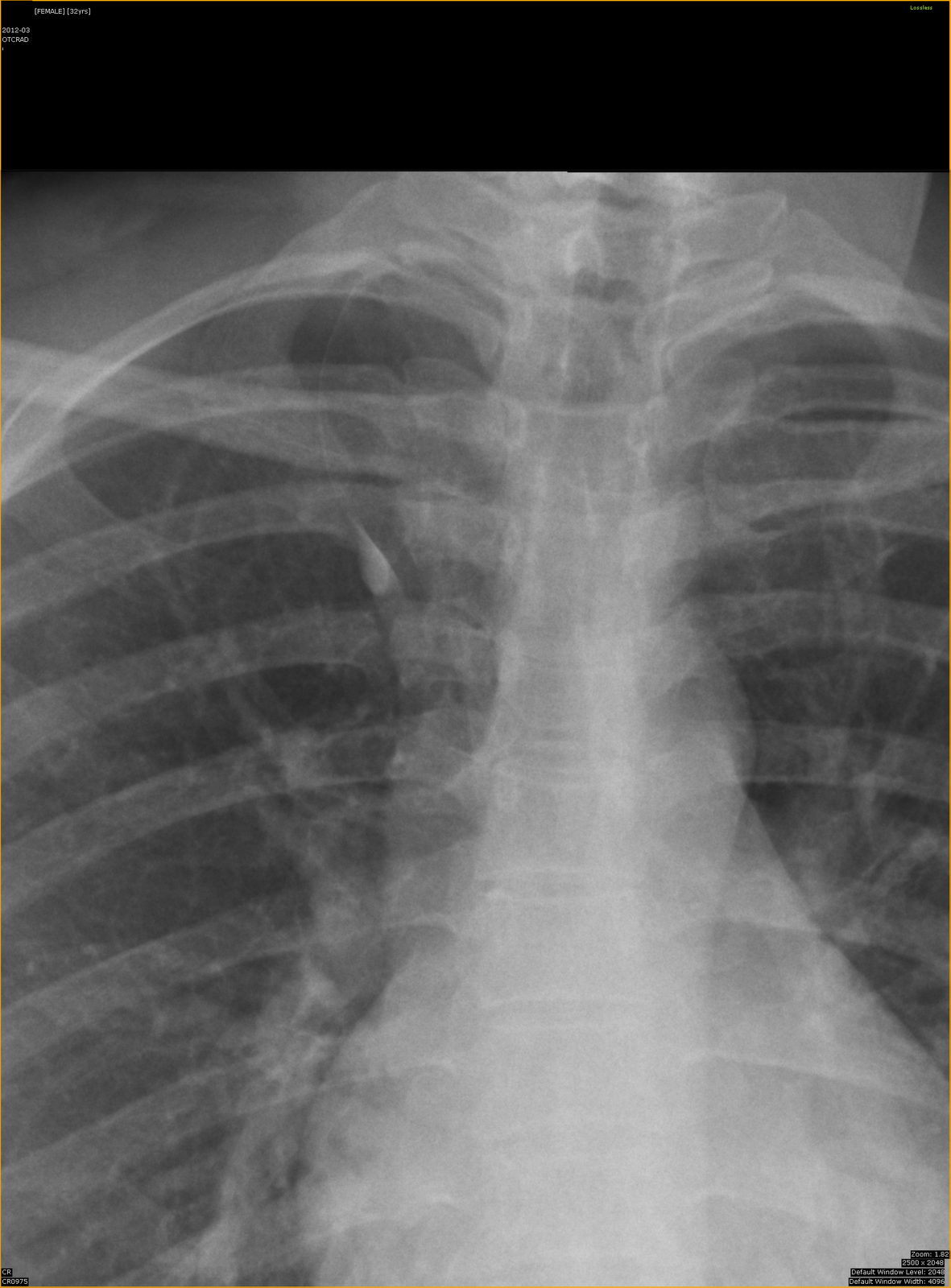
Skull radiographs showing numerous, discrete, well circumsribed small, lytic round lesions in the skull and mandible.

DIAGNOSIS: Multiple Myeloma

Discussion:

  • a hematologic disorder involving overproduction of a monoclonal immunoglobulin by neoplastic plasma cells. Tumor cells proliferate in the bone marrow and overexpress RANK ligand, which causes increased osteoclast activity and, ultimately, bone resorption.
  • most common primary malignant bone tumor in patients older than 40 years of age. 
  • primarily affects older people (only 2% of patients are younger than 40 years of age).
  • At least 30% cancellous bone loss is required to visualize an intramedullary destructive process with radiographs.
  • a disease of older patients. The disease can present with diffuse demineralization, which may be indistinguishable from the pattern found in patients with simple senile osteoporosis.
  • 2 sclerotic forms of myeloma:


  • A rare form, known as POEMS syndrome (polyneuropathy, organomegaly, endocrinopathy, monoclonal gammopathy, and skin changes), that may demonstrate sclerotic lesions on radiographs, but this condition is responsible for fewer than 1% of myeloma cases.
  •  Mixed lytic and sclerotic lesions.


  • Radiographs of treated myeloma lesions also may rarely show areas of abnormal bone architecture with sclerosis. Usually, little reactive bone sclerosis or periosteal reaction is seen (1)
  • If associated with bone marrow plasmacytosis and elevated blood gamma-globulins, the diagnosis of myeloma is certain.
  • False-positive examinations are encountered when multiple lytic lesions are found. In these patients, perform additional studies because the most likely source of this pattern is metastatic disease, not myeloma.
  • Diffuse osteopenia that is found on radiographs is often a source of false-negative examinations because a substantial amount of cancellous bone must be destroyed before an intramedullary lesion becomes visible radiographically.
  • A sclerotic form of multiple myeloma, sclerosing myelomatosis, which is the most severe variant. It is uncommon and associated with polyneuropathy, organomegaly, endocrinopathy, monoclonal gammopathy, and skin changes (POEMS) syndrome.
  • Imaging features:

1. Radiographs 

– typically presents as well-defined intramedullary lytic lesions, which are characteristically described as having a “punched out” appearance. The lesions can vary in size and can be focal or diffuse. 

– commonly affects the axial skeleton (vertebrae are the most common sites of involvement), but it can affect the proximal appendicular skeleton,

– it rarely affects the distal appendicular skeleton. 

– less commonly present with diffuse osteopenia, often with vertebral compression fracture(s).


2. MRI or PET/CT

– multifocal or diffuse infiltration of bone marrow, and an intramedullary soft-tissue mass is often visualized.


  • Differential diagnoses (for multiple focal lytic lesions)


  • Metastases
  • Leukemia
  • Primary lymphoma of bone.


  • Treatment typically involves chemotherapy and possibly autologous hematopoietic stem cell transplant.
  • Bisphosphonates are often used to prevent pathologic fractures.


References:

1.  Mulligan ME. Myeloma update. Semin Musculoskelet Radiol. Sep 2007;11(3):231-9.

2. Angtuaco EJ, Fassas AB, Walker R, Sethi R, Barlogie B. Multiple myeloma: Clinical review and diagnostic imaging. Radiology. 2004;231(1):11-23.

3. Kyle RA, Gertz MA, Witzig TE, et al. Review of 1027 patients with newly diagnosed multiple myeloma. Mayo Clin Proc. 2003;78(1):21-33.

4. Rothschild BM, Hershkovitz I, Dutour O. Clues potentially distinguishing lytic lesions of multiple myeloma from those of metastatic carcinoma. Am J Phys Anthropol. 1998;105(2):241-250.

5. Siegel RL, Miller KD, Jemal A. Cancer statistics,  2015. CA Cancer J Clin. 2015;65(1):5-29.

6. Dr. Neil Harrison, University of Pennsylvania Department of Radiology.

Liver (Biliary) Cystadenoma

CLINICAL DATA: Presented with upper abdominal discomfort and right chest discomfort for 3/12.

  • Normal LFT.
  • CA19-9 elevated (1556) = normal 0-37

MULTIPHASE CT LIVER STUDY:


Plain phase: A well defined cystic lesion in segment 1 LIVER with internal coarse calcifications. Largest internal calcifications measuring 1.7cm x 3.2cm.


Arterial phase: A well defined cystic lesion with attenuation of ~52HU in segment 1 liver.

Portal phase: The cystic lesion in segment 1 liver has attenuation of 33HU-36HU. It is slightly compressing the adjacent IVC. No hepatic / IVC vein thrombosis.

Portal phase: The C-C dimension of the S1 cystic lesion with internal calcification measuring 6.2cm. It is compressing the adjacent main portal vein but no thrombosis.

Portal phase. The S1 cystic liver lesion measuring 6.2cm x 5.1cm (axial dimension).

Delayed 3mins phase: Attenuation of the S1 cystic liver lesion of 35HU – 41HU. No ascites. No enlarged node in portal hepatic and para-aortic region. Thin peripheral enhancement.

Management: Surgical excision was performed. HPE Caudate lobe lesion of benign serous cystadenoma of the liver.

DISCUSSION:

Cystic lesions of the liver in the adult can be classified as developmental, neoplastic, inflammatory, or miscellaneous.

Developmental cystic liver lesions:

  • Hepatic cyst.
  • Polycystic liver disease.
  • Bile duct hamartoma.
  • Caroli disease.

Neoplastic cystic liver lesions:

  • Undifferentiated Embryonal Sarcoma
  • Biliary Cystadenoma and Cystadenocarcinoma
  • Cystic Subtypes of Primary Liver Neoplasms
  • Cystic Metastases

Inflammatory cystic liver lesions:

  • Liver abscess
  • Hepatic echinococcosis (intrahepatic hydatid cyst)

Miscellaneous cystic liver lesions:

  • Hepatic Extrapancreatic Pseudocyst
  • Hematoma
  • Biloma

Biliary (Hepatic) cystadenoma are rare, usually slow growing,  unilocular or multilocular cystic tumors that represent less than 5% of intrahepatic cystic masses of biliary origin (1,2,3).

  • In 1892, Keen reported the first case of hepatic cystadenoma which now accounts for 5% of all cystic lesions of the liver.
  • Age: >30 years (82%), peak incidence in 5th decade, M:F= 1:4
  • uncommon benign cystic neoplasm of the liver.
  • arise from the mucin-secreting bile duct epithelium and can grow quite large before exerting mass effect on adjacent structures and becoming symptomatic.
  • Two types of hepatic cystadenomas are described pathologically:
  • 1. mucinous  type –  the predominant type (95% of cases) that occurs in women
  • 2. serous type .
  • originates in the bile ducts and is lined by mucin-secreting columnar or cuboidal epithelium.
  • generally intrahepatic in 85%.
  •  among intrahepatic cystadenomas, 55% occur in the right lobe, 29% occur in the left lobe, and 16% occur in both lobes (2).
  • size varies in diameter from 1.5 to 35 cm.
  • occur predominantly in middle-aged women (5).
  • benign tumors, but they have a high rate of recurrence and a potential for neoplastic transformation in approximately 10% of cases.
  • considered premalignant lesions (3).
  • fluid within the tumor can be proteinaceous, mucinous, and occasionally gelatinous, purulent, or hemorrhagic due to trauma (2,3).
  • superinfection of the tumor may cause leukocytosis.
  • Carbohydrate antigen (CA) 19-9 levels may be elevated in some cases.
  • Carcinoembryonic antigen (CEA) and alpha-fetoprotein levels are usually normal.
  • no specific imaging features that permit reliable differentiation of biliary cystadenoma from biliary cystadenocarcinoma.
  • Complications of hepatic cystadenomas include the following:
    • Bleeding
    • Rupture
    • Obstructive jaundice
    • Malignant transformation
    • Infection
    • gastric outlet obstruction
    • ascites
    • Inferior vena cava obstruction
  • Ultrasound:
  1. appears as a unilocular or multilocular cyst with enhanced through transmission. The content of the cysts may range from completely anechoic to having low-level echoes from blood products, mucin, or proteinaceous fluid.
  2. Mural nodules and papillary projections may project into the cyst lumen.
  3. If septal or wall calcification is present then acoustic shadowing may be seen.
  4. more reliable in correctly diagnosing the degree of septation, which is a predominant feature of both cystadenomas and cystadenocarcinomas.
  • CT:
  1. as a solitary cystic mass with a well-defined thick fibrous capsule, mural nodules, internal septa, and rarely capsular calcification (2,3). Uni- or multilocular cystic mass (with unilocular being far more prevalent).
  2. polypoid, pedunculated excrescences are seen more commonly in biliary cystadenocarcinoma than in cystadenoma, although papillary areas and polypoid projections have been reported in cystadenomas without frank malignancy (4).
  3. can range from that of water (HU = 0) to quite hyperattenuating if the cyst has been complicated by recent hemorrhage.
  4. calcifications of septa or cyst wall may be seen.
  5. the septa may enhance following administration of contrast.
  6. occasional mural nodules.
  7. In published studies, CT scanning was less sensitive in correctly identifying septa within a cystic lesion (6,7,8)
  • MRI:
  1. uncomplicated biliary cystadenoma correlate well with the pathologic features: The appearance of the content is typical for a fluid-containing multilocular mass, with homogeneous low signal intensity on T1-weighted images and homogeneous high signal intensity on T2-weighted images.
  2. Variable signal intensities on both T1- and T2-weighted images depend on the presence of solid components, hemorrhage, and protein content (2,3).
  • may recur after excision and have potential to develop into biliary cystadenocarcinoma.
  • the prognosis of hepatic cystadenomas is extremely good if patients undergo a complete surgical resection

References:

1. Murphy BJ, Casillas J, Ros PR, et al. The CT appearance of cystic masses of the liver. RadioGraphics 1989; 9:307-322

2.  Palacios E, Shannon M, Solomon C, et al. Biliary cystadenoma: ultrasound, CT, and MRI. Gastrointest Radiol 1990; 15:313-316
  1. Buetow PC, Midkiff RB. Primary malignant neoplasms in the adult.Magn Reson Imaging Clin N Am 1997; 5:289-318
  2. Powers C, Ros PR, Stoupis C, et al. Primary liver neoplasms: MR imaging with pathologic correlation.RadioGraphics 1994; 14:459-482. 
  3. Levy AD, Murakata LA, Abbott RM et-al. From the archives of the AFIP. Benign tumors and tumorlike lesions of the gallbladder and extrahepatic bile ducts: radiologic-pathologic correlation. Armed Forces Institute of Pathology. Radiographics. 22 (2): 387-413. 
  4. Korobkin M, Stephens DH, Lee JK, et al. Biliary cystadenoma and cystadenocarcinoma: CT and sonographic findings. AJR Am J Roentgenol. 1989 Sep. 153(3):507-11.
  5. Eisenberg RL. Ultrasound and CT of gallbladder, liver and spleen. Gastrointestinal Radiology: A Pattern Approach. Philadelphia, Pa: Lippincott-Raven. 1996:1085-7.
  6. Colon-Negron E, Meyer CA, Halvorsen RA. Liver imaging: surgical segmental anatomy. In: Taveras JM, Ferrucci JT, eds. Radiology: Diagnosis, Imaging, Intervention. Philadelphia, Pa: Lippincott Williams & Wilkins; 1996. Vol 4: 1-10.
  7. 9. Mortelé KJ, Ros PR. Cystic focal liver lesions in the adult: Differential CT and MR imaging features. Radiographics. 2001;21(4):895-910.

    10. Roth CG, Deshmukh S. Fundamentals of Body MRI. 2nd ed. Philadelphia, PA: Elsevier; 2017.

    11. Siegelman E. Body MRI. Philadelphia, PA: Elsevier-Saunders; 2005.