Ig G4 Related Diseases

CLINICAL DATA:
A 47-year-old man presented with worsening bilateral orbital swelling, parotid swelling and submandibular swelling for several months.
ESR 90mm/hr

Creatinine 150umol/l

Elevated serum uric acid

IMAGING INVESTIGATIONS:

  • Bilateral swelling of the lacrimal glands. Homogenous enhancement of the involved glands with no liquefaction/calcification.

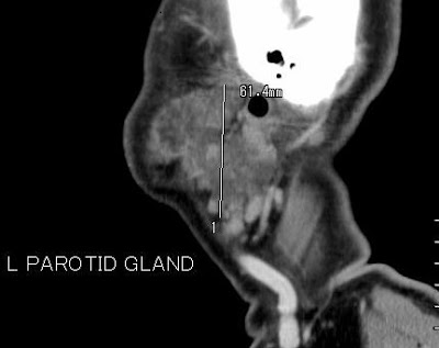
  • Bilateral swelling of parotid glands. Heterogenous enhancement predominantly in the periphery of the glands. No liquefaction/calcification.
  • No enlarged neck node.

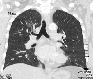
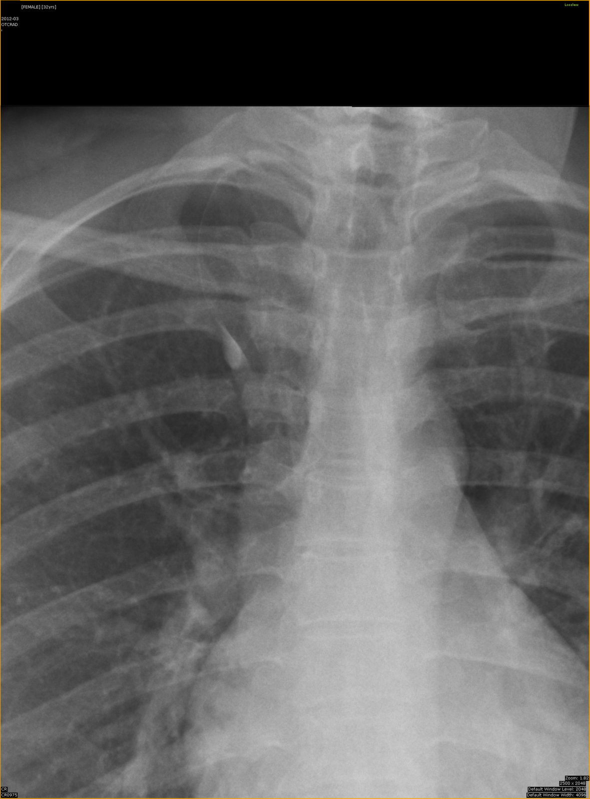
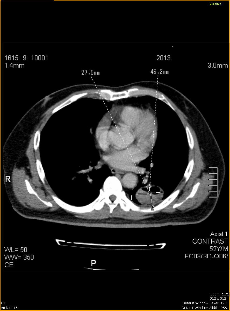
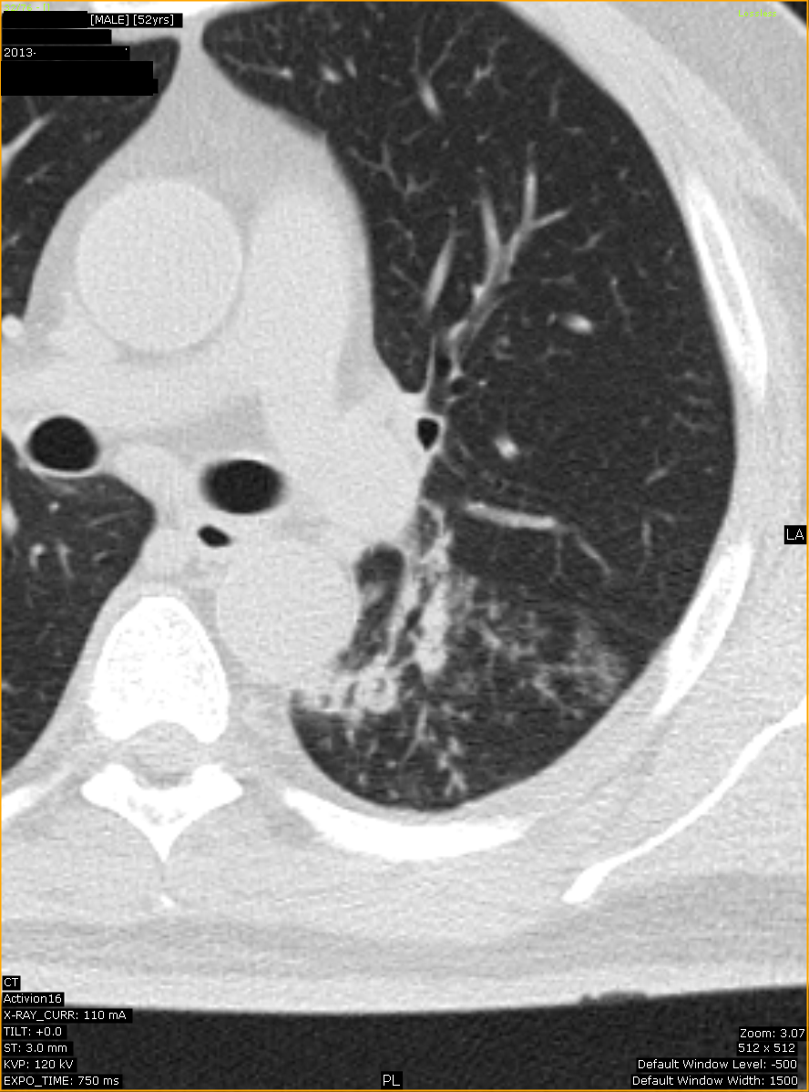
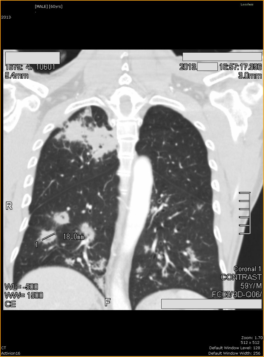
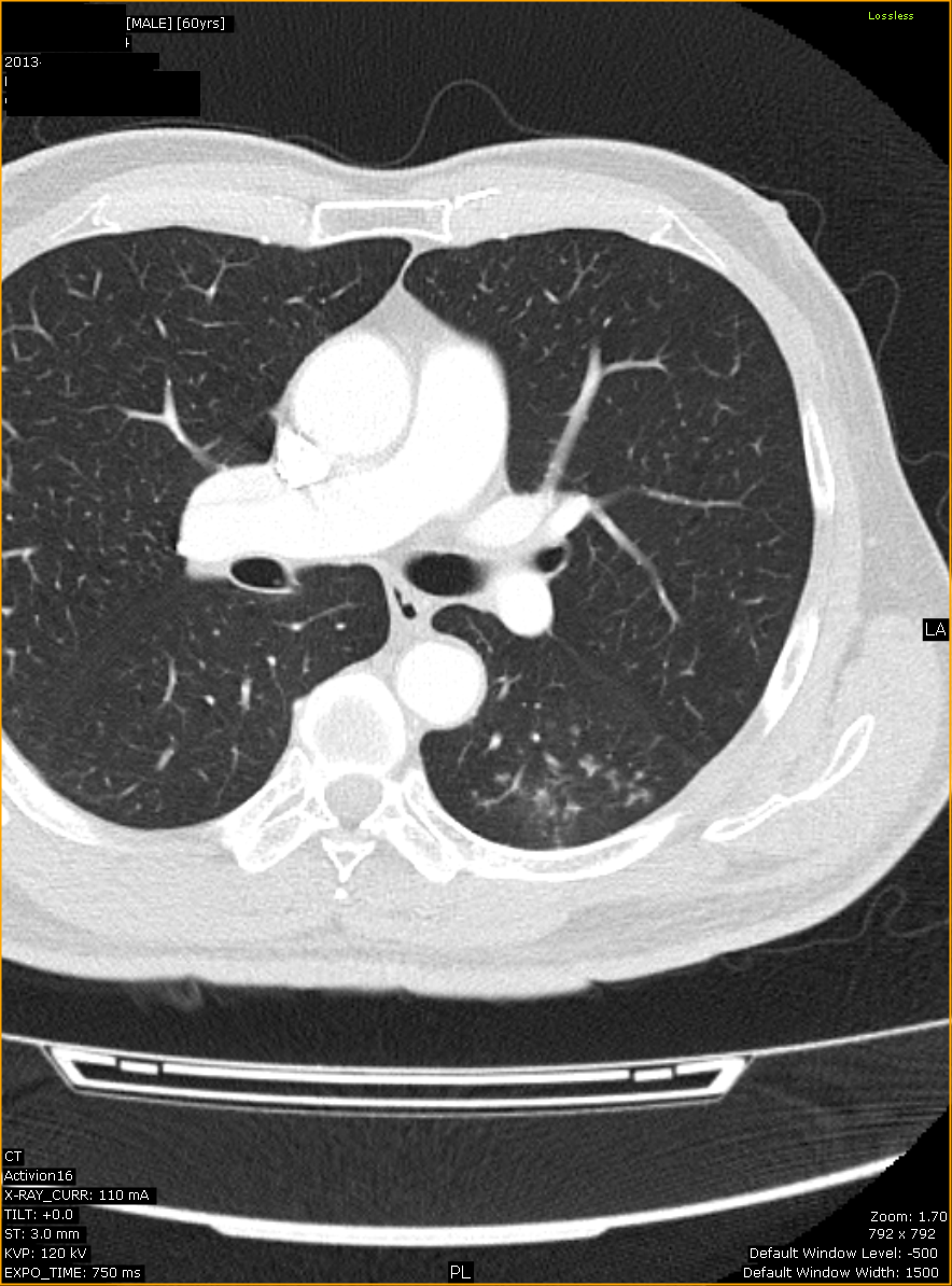
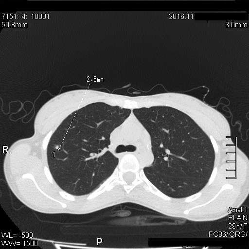
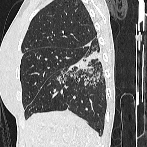
  • Focal ground glass opacity in the right upper lobe.

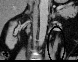
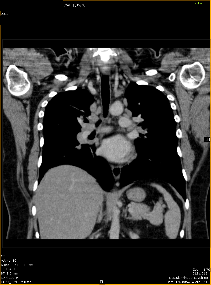
  • Ureteric wall thickening of the visualized upper left ureter.
  • Mild left hydronephrosis.
DIFFERENTIAL DIAGNOSIS:
1. Mikulicz Syndrome –  a form of Sjögren syndrome (type 1). However more recently it is considered in IgG4 related disease spectrum.
BIOPSY OF THE SUBMANDIBULAR GLAND WITH STAINING: Ig G 4 disease.
DISCUSSION:
  • characterized by fibro-inflammatory lesions rich in IgG4-positive plasma cells and, often but not always, elevated serum IgG4 concentrations.
  • the pathogenesis of IgG4-related disease is poorly understood, but there are findings consistent with both an autoimmune and an allergic disorder.
  • was recognized as a systemic disease in 2003, when extrapancreatic manifestations were identified in patients with autoimmune pancreatitis. The pancreas is the most commonly affected organ in IgG4-related disease.
  • affecting virtually every organ system and has been identified in the biliary tree, salivary and lacrimal glands, periorbital tissues, lungs, lymph nodes, thyroid gland, kidneys, prostate gland, testicles, breasts, skin, meninges, retroperitonrum, mesentery, and pituitary gland (1-3).
  • Extrapancreatic lesions may sometimes develop before or up to 15 years after pancreatitis.
  • imaging plays an important role in demonstrating infiltration and enlargement of involved organs.
  • Immunohistochemical staining for IgG4 is required. The presence of IgG4-positive plasma cells is required for diagnosis of IgG4-related disease, but some IgG4-positive plasma cells can also be detected in other inflammatory diseases. However, it has been suggested that dense infiltrates of IgG4-plasma cells (>10–50 per high-power microscopic field, depending on the affected organ) are highly specific for IgG4-related disease (3). A ratio of IgG4-positive to IgG-positive plasma cells higher than 40% is also helpful in distinguishing between IgG4-related disease and non–IgG4-related inflammatory conditions (3-5).
  • Elevated serum IgG4 concentration is common in but not specific for IgG4-related disease (6).
  • Serum IgG4 concentration may be normal in 20%–40% of cases of biopsy-proved IgG4-related disease (3, 7,8). Thus, neither an increase in serum IgG4 concentration nor an elevated number of IgG4-positive plasma cells in tissue is specific for IgG4-related disease (18).
  • Response to steroid therapy is excellent in most patients, although some patients are refractory to such therapy (7).
IgG4-related pancreatitis:
  • Nearly 40% of patients with IgG4-related pancreatitis also have salivary and/or lacrimal gland involvement, which is characterized by bilateral and painless swelling of the glands (9).This manifestation may precede or accompany pancreatitis, but it may also occur in isolation.
  • The pancreas is the most common site affected, followed by the biliary tree and kidneys, but virtually any site can be involved, including the gallbladder, lymph nodes, retroperitoneum, mesentery, lungs, breast, prostate, skin, and a variety of areas in the head and neck.
IgG4-related lacrimal/salivary glands:
  • Recent reports suggest that a substantial proportion of idiopathic orbital inflammations—also called orbital pseudotumors—are associated with IgG4-related disease(10).
IgG4-related renal involvement;
  • Renal involvement may be present in up to 35% of patients with autoimmune pancreatitis (11,12). The presence of renal lesions in patients with pancreatic disease has been used to help differentiate autoimmune pancreatitis from pancreatic cancer, since their presence strongly suggests the former condition. Five patterns of disease have been described:
  1. bilateral round or wedge-shaped peripheral cortical lesions (the most common)
  2. diffuse patchy involvement
  3. a rim of soft tissue around the kidney,
  4. bilateral nodules in the renal sinuses, and diffuse wall thickening of the renal pelvis (11).
  •  Patients with IgG4-related nephritis have no hematuria and usually improve after undergoing corticosteroid therapy.
Ig G4-related pleural/lung involvement:
  • Four major types of IgG4-related pulmonary disease have been defined:
  • (a) a type characterized by solid nodular or mass like lesions,
  • (b) a type characterized by round ground-glass opacities,
  • (c) alveolar interstitial disease, and
  • (d) bronchovascular disease (13).
  • When IgG4-related lung disease appears as a round ground-glass opacity at CT, imaging findings may suggest bronchoalveolar carcinoma.
  • Visceral or parietal pleural thickening has been reported, sometimes involving the subpleural lung parenchyma
References:
1. Zen Y, Harada K, Sasaki M, et al. IgG4-related sclerosing cholangitis with and without hepatic inflammatory pseudotumor, and sclerosing pancreatitis-associated sclerosing cholangitis: do they belong to a spectrum of sclerosing pancreatitis? Am J Surg Pathol 2004;28(9):1193–1203.
2. Hennessey JV. Riedel’s thyroiditis: a clinical review. J Clin Endocrinol Metab 2011;96(10):3031–3041.
3. Deshpande V, Zen Y, Chan JK, et al. Consensus statement on the pathology of IgG4-related disease. Mod Pathol 2012;25(9):1181–1192.
4. Umehara H, Okazaki K, Masaki Y, et al. Comprehensive diagnostic criteria for IgG4-related disease (IgG4-RD), 2011. Mod Rheumatol 2012;22(1):21–30.
5. Zen Y, Nakanuma YIgG4-related disease: a cross-sectional study of 114 cases. Am J Surg Patho 2010; 34(12): 1812-1819.
6. Aalberse RC, Stapel SO, Schuurman J, Rispens T. Immunoglobulin G4: an odd antibody. Clin Exp Allergy 2009;39(4):469–477
7. Stone JH, Zen Y, Deshpande V. IgG4-related disease. N Engl J Med 2012;366(6):539–551.
8. Sah RP, Chari ST. Serologic issues in IgG4-related systemic disease and autoimmune pancreatitis. Curr Opin Rheumatol 2011;23(1):108–113.
9.  Hamano H, Arakura N, Muraki T, Ozaki Y, Kiyosawa K, Kawa S. Prevalence and distribution of extrapancreatic lesions complicating autoimmune pancreatitis. J Gastroenterol 2006;41(12):1197–1205.
10.  Wallace ZS, Khosroshahi A, Jakobiec FA, et al. IgG4-related systemic disease as a cause of “idiopathic” orbital inflammation, including orbital myositis, and trigeminal nerve involvement. Surv Ophthalmol 2012;57(1):26–33.
11. Takahashi N, Kawashima A, Fletcher JG, Chari ST. Renal involvement in patients with autoimmune pancreatitis: CT and MR imaging findings. Radiology 2007;242(3)
12.  Khalili K, Doyle DJ, Chawla TP, Hanbidge AE. Renal cortical lesions in patients with autoimmune pancreatitis: a clue to differentiation from pancreatic malignancy. Eur J Radiol 2008;67(2):329–335.
13. Inoue D, Zen Y, Abo H, et al. Immunoglobulin G4-related lung disease: CT findings with pathologic correlations. Radiology 2009;251(1):260–270

Chest findings in scleroderma

A 73 year old man with underlying scleroderma presenting with progressive shortness of breath.
A Chest X-Ray was performed.

  • Fibrotic changes involving the lower two thirds of the lung, with associated volume loss and honeycombing.
  • Ellevation of diaphragms suggestive of restrictive lung disease.
  • Subpleural reticular or reticulonodular opacity.
  • Cardiomegaly.

DISCUSSION:

Plain film:

  • insensitive to early changes
  • may be normal despite respiratory function test abnormalities.
  • pulmonary fibrosis 
  • dilated oesophagus
  • eggshell calcification of mediastinal nodes
  • pleural effusions are uncommon
  • enlargement of cardiac silhouette and pulmonary arteries due to scleroderma induced pulmonary vascular disease may also be evident. 

HRCT Thorax:

Azygous fissure


Clinical data: A foreign worker came for routine medical examination.

CHEST RADIOGRAPH (PA ERECT):


Findings:

  • Accessory fissure forming a thin ‘inverted comma’ line.
  • Azygous vein forming the head of a ‘tadpole’.

Disccusion:

    • an accessory fissure occurs in approximately 1-2% of individuals.
    • It is typically an incidental radiographic finding.
    • It has no associated signs or symptoms.
    • No treatment is necessary, as this is a normal anatomic variant.
    • vein appears to run within the lung wrapped by four layers of pleura (two parietal layers and two visceral layers) giving the appearance of ‘head of a tadpole’.
    • the azygous vein has a more lateral course and descends along a fissure in the right upper lobe, resulting in azygous fissure.
    • The azygos vein and trigone also are often seen.
      • Azygos vein: Appears as an ovoid, tear-shaped opacity in the inferior aspect of the accessory fissure.
      • Trigone: Appears as a triangular opacity that marks the superior aspect of azygos fissure.
    • accessory fissures of the lung usually occur at the borders of bronchopulmonary segments. They are normal variants.
    • other accessory fissures include inferior accessory fissure,  superior accessory fissure and left minor fissure.
    • the inferior accessory fissure surrounds the medial basal segment of the lower lobe.
    • the superior accessory fissure seperates the superior segment of lower lobe from basal segments.
    • left minor fissure separates the lingula from the rest of the left upper lobe. 
    • accessory fissures, like all other fissures, serve not only as natural barriers against infection but also help in localizing any focal pulmonary parenchymal disease.
  • Major fissures are oblique fissures and right transverse fissure.
  • The left lung is divided into two lobes, upper and lower. These lobes have their own pleural covering and these lie together to form the oblique (major) fissure.
  • The right lung, there is an oblique fissure and a horizontal fissure, separating the lung into three lobes – upper, middle, and lower. 
  • The horizontal fissure (right) is often seen on a normal frontal view CXR whereas the oblique fissures are often seen on a normal lateral view

CT THORAX:

Pectus Excavatum

ROUTINE MEDICAL SCREENING:

CHEST RADIOGRAPH (PA ERECT VIEW)

  • poor delineation of right heart border.
  • increased density of the inferomedial lung field.
  • horizontal posterior ribs.
  • vertical anterior ribs.
  • displacement of heart towards the left.

On examination, the above patient has pectus excavatum.

 DISCUSSION:
  • also known as funnel chest.
  • congenital chest wall deformity characterised by concave depression of the sternum resulting in radiographic alterations.
  • the sternum is depressed inward and the ribs protrude anteriorly.
  • most common chest wall deformity.
  • occurs in up to 1 in 300-1000 births (1,2).
  • occasionally associated with:
  • 1. mitral valve prolapsed.
  • 2. restrictive pulmonary function test abnormalities.
  • 3. cardiac functional abnormalities.
  • Differential diagnosis: can be troublesome on frontal chest radiographs. 
  • right middle lobe consolidation/atelectasis.
  • left para-aortic soft tissue density:
  • a. lung or paravertebral mass.
  • b. atelectasis/consolidation of the medial segment of the left lower lobe.
  • mediastinal mass due to deformation of the cardiomediastinal contour.
  • The diagnosis is obvious on lateral projection, and of course cross sectional imaging.

References:

1. Dähnert W. Radiology review manual. Lippincott Williams & Wilkins. (2007) ISBN:0781738954. 

2. Kelly RE, Cash TF, Shamberger RC et-al. Surgical repair of pectus excavatum markedly improves body image and perceived ability for physical activity: multicenter study. Pediatrics. 2008;122 (6): 1218-22. 

Sarcoidosis

CASE 1


Clinical data:

A 36 years old Indian man was admitted for uncontrolled hypertension. History of hypertension since 4 years ago.

Having recurrent dry cough for more than 1 year. No fever.

CHEST RADIOGRAPH:

 
  • CXR showed hilar opacity.
 CT THORAX:

  • Coronal images showing multiple enlarged discrete solid mediastinal nodes. No lung parenchymal abnormality. Enlarged right paratracheal and bilateral hilar nodes constituting the ‘1-2-3 sign’ (Garland triad).

  • Enlarged discrete solid lymph nodes in pretrachea, paratrachea, tracheo-bronchial, hilar and aorto-pulmonary window.

BIOPSY: Sarcoidosis.

CASE 2

CLINICAL DATA: A 51-year old Indian man presented with weight loss and abdominal discomfort. Strong history of alcohol consumption.

CT STUDY:

  • Hepatosplenomegaly with nodular appearance in both these organs. Nodular liver outline.
  • Portal vein and splenic vein diameter is within normal limit. No dilated collaterals at splenic hilum.
  • Enlarged discrete para-aortic nodes below the origin of renal arteries.

  • Enlarged right inguinal node.

CHEST RADIOGRAPH:

  • Nodular opacities in both upper and mid lung fields (right > left).
  • Peribronchial cuffing in the right upper and mid lung field.
  • No widening of the mediastinum. No pleural effusion.

HRCT THORAX (MIP IMAGES):

  • Ill defined small nodules scattered predominantly in both upper lobes and right middle lobe. These nodules have stellate appearance. Some nodules are miliary-like.
  • Nodular peribronchial interstitium (perilymphatic distribution).

  • No enlarged mediastinal node.
  • Discrete enlarged nodes in bilateral axillary regions.

Excision biopsy of right inguinal node: HPE revealed SARCOIDOSIS.

 Discussion:
  • a multisystem inflammatory disease of unknown etiology that predominantly affects the lungs and intrathoracic lymph nodes. 
  • presence of noncaseating granulomas (NCGs) in affected organ tissues.
  • In USA, the incidence ranges from 5-40 cases per 100,000 population.  
  • Internationally, the incidence is 20 cases per 100,000 population in Sweden and 1.3 cases per 100,000 population in Japan.
  • The disease remains hidden and often is misdiagnosed as tuberculosis.
  • 5-10% of patients, the chest radiograph is normal.
  •  Bilateral hilar lymphadenopathy is the most common radiographic finding. Pulmonary sarcoidosis may manifest with various radiologic patterns: Bilateral hilar lymph node enlargement is the most common finding, followed by interstitial lung disease.
  • The clinical staging of sarcoidosis is based on the pattern of chest radiographic findings:
  • Stage 0 is a normal chest radiograph.
  • Stage I is lymphadenopathy only.
  • Stage II is lymphadenopathy and lung parenchymal disease.
  • Stage III is parenchymal lung disease only.
  • Stage IV is pulmonary fibrosis. It is often associated with upper lung fibrosis with other findings which include honeycombing, bullae and cyst formation, and bronchiectasis. The fibrosis may result in abnormal central conglomeration of hilar and perihilar structures, and upper lobe conglomerate masses.
  • radiologic staging does not correlate well with the severity of pulmonary function abnormalities.
  • CT is more sensitive than radiography in the detection of lymphadenopathy and subtle parenchymal disease. HRCT results may be normal in the presence of microscopic disease.
  • Isolated unilateral hilar lymphadenopathy is an unusual manifestation of sarcoidosis, occurring in only 1-3% of patients.
  • Mediastinal lymphadenopathy with no associated hilar lymphadenopathy or unilateral hilar lymphadenopathy occurs more frequently in such older patients.
  • Adenopathy usually decreases as parenchymal disease increases. In stage III disease, intrathoracic adenopathy does not develop subsequent to parenchymal disease.  If adenopathy develops, think of lymphoma or TB.
  • Hilar and/or mediastinal lymphadenopathy is not specific for sarcoidosis, and similar findings may occur in lymphoma, leukemia, metastases, and fungal and viral infections.
  • At high-resolution CT, the most typical findings of pulmonary involvement are micronodules with a perilymphatic distribution, fibrotic changes, and bilateral perihilar opacities (1).
  • Atypical manifestations, such as mass-like or alveolar opacities, honeycomb-like cysts, miliary opacities, mosaic attenuation, tracheobronchial involvement, and pleural disease, and complications such as aspergillomas (1).
  • Patterns of lung disease: Reticulonodular (46%), Acinar pattern (20%) and Larger nodules which include “Alveolar sarcoid” (2%), coalescence of numerous interstitial granulomas and air bronchograms present.
  •  DDX
  • o       Alveolar cell ca
  • o       Alveolar proteinosis
  • o       Lymphoma.
  • HRCT findings include:

    a. areas of ground-glass attenuation. The foci of ground-glass attenuation may represent areas of active alveolitis or diffuse microscopic interstitial granulomas.

    b. subpleural nodules. The nodules, which correspond to coalescent interstitial granulomas, have irregular margins.

    c. Perivascular nodules which appear as beading and irregular thickening of bronchovascular bundles and thickening of interlobular septa.

Varicella-Zoster Pneumonia

CLINICAL DATA: Day 3 chicken pox complicated with cough.


 Day 1 admission CXR. Consolidation in retrocardiac region.


Day 3 admission. 

  • Patchy diffuse air-space consolidation predominantly near hila and lung bases.
  • Widespread ill-defined nodular opacities (acinar nodular pattern).


 Day 7 admission. 

  • Improved. No lung scarring.


DISCUSSION:


Varicella-zoster virus (VZV) most commonly is a self-limited benign disease (chickenpox) in children. However, in adults it tends to cause significant complications such as VZV pneumonia. 

2 types of pathologic reactions and radiologic aspects can be observed:

             (1) usual, long-standing, or insidious course of pneumonia; and

             (2) rapidly progressive or virulent pneumonia.

Radiological changes:

  • radiographic manifestations usually appear 2-5 days after the rash does.
  • tend to clear in 3-5 days in mild disease and take up to several weeks or months to clear in widespread disease.
  • Patchy, diffuse air space consolidation
  • Tendency to coalesce near hila
  • Widespread nodules can occur (30%) appearing as ill-defined, 5- to 15-mm nodular opacities (acinar nodular pattern). The nodules are seen in the lung periphery (bases), coalescing near the hila;
  • Tiny calcifications remain in 2% (DDX is histoplasmosis, alveolar microlithiasis).
  • CT images may show nodules with a surrounding halo of ground-glass opacity, patchy ground-glass opacity, and coalescence of nodules.
  • Unique complication consists of the late appearance (years after onset of pneumonia) of 2- to 3-mm dense calcifications, which are well defined, scattered, and predominant in the lower half of the lungs. 

11% mortality rate.

Pneumonia due to bacterial superinfection is segmental in distribution, it affects 1 or both lower lobes, and it is frequently associated with atelectasis. The presence of a dense opacity is more suggestive of a bacterial etiology (88%) than a viral etiology (36%). 


Round Pneumonia

CLINICAL DATA: One year old child with high grade fever, productive cough and leucocytosis.

CHEST RADIOGRAPHS:

Round pneumonia in the right upper lobe (day 1 admission).

Follow up CXR 10 days later.


DISCUSSION:

Spherical pneumonia caused by:

  • Haemophilus influenzae
  • Streptococcus
  • Pneumococcus.

Location:

  • Usually lower lobe
  • Most often posterior

mean age of patients with round pneumonia is 5 years. 90% of patients who present with round pneumonia are younger than twelve (1).
majority of cases (98%), they are solitary (1).
may have slightly irregular border and contain air bronchogram


References:

1. Kim YW, Donnelly LF. Round pneumonia: imaging findings in a large series of children. Pediatr Radiol. 2007;37 (12): 1235-40

Pulmonary Tuberculosis

CASE 1

CLINICAL DATA:

A man presented with chronic cough. Occasional haemoptysis. No previous illness before.

Mantoux test – indurated area of 24mm.

CHEST RADIOGRAPH:

  • Small cavitations in the left upper lobe with fibrosis and nodular opacities.

HRCT THORAX:

Fibrosis and small lung cavitations in the left upper lobe.


CASE 2:

A 47 year-old man with chronic cough. Poorly controlled DM status.

Chest radiograph:

  • Cavitating pneumonia of left upper lobe.

Sputum AFB direct smear is positive.

CASE 3

CLINICAL DATA: A 29 year-old lady with persistent cough for 1month. Delivered a baby 3 month ago. History of contact with pulmonary TB.

CHEST RADIOGRAPH:

Reticulo-nodular opacities in both upper lobes (right > left) with minimal fibrosis in right lung apex.

Sputum AFB: positive.

CASE 4

CLINICAL DATA:

A 52 years-old gentleman with underlying DM. Presented with cough and fever for 2months duration. Admitted to hospital for haemoptysis.

Previous screening CT THORAX 2 years ago showed a homogenous solid mass in superior segment of left lower lobe presuming to be lung hamartoma (images not available).

HbA1C: 11.6%

ESR 57mm/hour.

CHEST RADIOGRAPH:

  • A cavitating lung lesion in the superior segment of left lower lobe.

CONTRAST ENHANCED CT THORAX:

The previously seen solid lung lesion (presume to be lung hamartoma) in superior segment of left lower lobe showed cavitation. It is measuring 4.6cm x 2.7cm in size.This lesion is representing lung tuberculoma.

There is tree-in-buds appearance adjacent to the cavitating lung lesion.

SPUTUM: AFB 3+

CASE 5

CLINICAL DATA: A 60 years old man of poorly controlled diabetis mellitus presented with cough.No fever. No loss of appetite or loss of weight.

CHEST RADIOGRAPH:

  • Multiple lung nodules of varying sizes predominantly in lower and mid lung fields.
  • Patchy consolidation in the right upper lobe.

CT THORAX:

  • Lung consolidation in right upper lobe.
  • Cavitating lung lesion in left upper lobe.
  • Nodular opacities in both lower lobes.

  • Tree-in-buds in superior segment of left lower lobe.

Spiculated multiple lung nodules in both lower lobes. Some appearing as flame-shaped lung lesions.

SPUTUM: AFB 3+.


CASE 6

CLINICAL DATA: A 14-years old girl presented with chronic cough. Patient is underweight and pale looking. Enlarged left neck, left supraclavicular and left axillary nodes.

CHEST X-RAY:

  • Consolidation of left lung with nodular opacities.
  • Left hydropneumothorax.

CT THORAX:

  • Cavitation consolidation of left lung. Lung abscess in left lower lobe. Left hydronpneumothorax.
  • Patchy consolidation in right upper lobe with tree-in-buds appearance.

  • Peripheral enhancing with low center density enlarged nodes in the left neck, left supravlavicular, left axillary region and mediastinum.
  • Cavitation lung consolidation in left lung.

  • Lung abscess in left lower lobe.
  • Left hydropneumothorax.

SPUTUM: AFB strongly positive.


CASE 7

CLINICAL DATA: A 29-yr-old lady presented with shortness of breath and hoarseness of voice.

Total White – normal.

ESR 26mm/hr (1-20)

Weight: 35.8  Kg

INVESTIGATIONS:

CHEST X-RAY:

  • Nodular consolidation in bilateral lung fields.
  • Thickened right lung fissure.
 HRCT THORAX:
  • A non spiculated lung nodule in the right upper lobe favouring lung granuloma.

  • Tree-in-buds involving predominantly superior segment right upper lobe and anterior-lingular segment left lower lobe.
  • Thickened adjacent lung fissures.
  • Patchy lung consolidation.

TB quantiferon posiitive.

Sputum culture: Mycobactrium TB positive.

DISCUSSION:

Types of TB infection:


1. Primary TB .

    • lungs are the primary organ of spread- accounting for about 70% of cases (1).
    • extrapulmonary infection generally occurs as a result of hematogenous dissemination from a clinically occult pulmonary focus (1).
    • typically a self-limited infection. 
    • about 5% of adults and up to 60% of infected children are asymptomatic (2).
    • in a less competent host, the infection is walled off, but the bacillus remains viable, but dormant for many years (3).
    • typically presents as a segmental or lobar consolidation usually involving the lower lobes (although any lobe may be involved) and the appearance is often indistinguishable from bacterial pneumonia (4).
    • Inhaled bacteria are deposited in the mid and/or lower lung. The bacteria then undergo intracellular multiplication with lymphatic and hematogenous spread. Bacteria survive in areas of the lung with high oxygen content.
    • multifocal involvement is seen in 12-24% of cases (5).
    • prevalence of lymphadenopathy is greatest in the pediatric age group (about 90-96% of affected children (4,6,7) and is seen in about 43% of adults (4).
    • Lymph node enlargement is a common finding with primary tuberculosis. This may occur with or without an upper lobe granuloma.
  • Pleural effusion is found in up to 40% of adults, but only 5-10% of children with primary infection (7).  Pleural fluid cultures are positive in only 20-40% of cases (pleural biopsy cultures are positive in 65-75% of cases) (5). Pleural effusion can be the only radiographic finding indicative of primary TB infection in about 5% of cases (5,7). 
  • Regression of radiographic findings is a slow process- requiring from 6 months to 2 years for resolution. 
  • Radiographic differentiation between active and inactive disease can only be made reliably on the basis of temporal evolution. The American Tuberculosis Association requires that a radiograph remain unchanged for a period of 6 months to indicate stable/inactive disease (8).
  • Computed tomography can detect the presence of adenopathy, parenchymal consolidations, or evidence of endobronchial spread not seen on plain film radiographs.  A normal chest radiograph has a high negative predictive value for the presence of active TB (5).
  • Common findings of infection in infants include mediastinal and hilar adenopathy (seen in 90-95% of cases (3).  The adenopathy is usually unilateral and located in the hilum or paratracheal region (3). On CT the nodes demonstrate central necrosis with rim enhancement (3).
  • Pulmonary tuberculosis can manifest as pulmonary nodules mimicking lung metastasis.

2. Miliary (disseminated) TB.

  • Typical miliary lesions may not be visible for 3 to 6 weeks after hematogenous dissemination (9). 
  • CXR reveals micronodular densities (1-2mm) diffusely throughout both lungs.
  • HRCT demonstrates a combination of sharp and poorly defined 1 to 3 mm nodules distributed throughout the lungs and have no relationship to the airways in their distribution. The nodules usually resolve within 2-6 months with treatment (4).

3. Progressive primary TB.

  • Widespread pulmonary infection due to impaired host immunity.

4. Reactivation or post-primary TB.

  • Reactivation infection usually develops in the apical/posterior segments of the upper lobes (83-85% of cases) or superior segment of the lower lobes (11-14% of cases) (2).
  • Patchy alveolar infiltrate. The cavities typically have thick, irregular walls which become smooth and thin with successful treatment (4).
  • Hilar or mediastinal adenopathy is unusual in reactivation TB.
  • An effusion may be the sole manifestation of reactivation TB (3).
  • In addition to an upper lobe granuloma, there are also typically satellite nodules which may coalesce and be able to be identified on chest x-ray. 
  • Tree-in-bud pattern opacities are another finding indicative of postprimary tuberculosis.
  • Cavitation is indicative of active and transmissible disease.

5. Tuberculous airway disease

  • CT of the chest during active infection will reveal irregular tracheobronchial narrowing and wall enhancement with I.V. contrast. The mediastinal fat around the trachea often demonstrates increased density consistent with inflammation. 

6. Chronic tuberculous empyema

  • On CXR there is usually a moderate to large loculated pleural fluid collection with pleural calcification and enlargement of the overlying ribs. 
  • CT demonstrates the loculated pleural fluid surrounded by a thick, calcified pleural rind (10).

7. Tuberculoma.

  • Well defined or have irregular margins and mimic a lung neoplasm.
  • Most lesions are less than 3 cm in size and calcification can be seen in 20-30% of cases (usually ndoular or diffuse). 
  • Small satellite nodules about the larger lesion can be found in up to 80% of cases.
  • Tuberculoma seems to be round or polygonal shape and primary lung cancer is more likely to be lobulated shape. The smooth border nodule is found only in tuberculoma (27%) whereas 93% of primary lung cancer had spiculated border compared to 73% among tuberculoma (p < 0.05). 

TB lymphadenitis:

  • In patients with active tuberculosis, nodes larger than 2 cm in diameter commonly show central areas of low attenuation on contrast-enhanced CT, with peripheral rim enhancement
  • The areas of relative low attenuation are not of water density, but range from about 40 to 60 HU; they are usually visible only on contrast-enhanced scans.

Tree-in-buds on HRCT LUNG:

  • Tree-in-buds apperance in the lungs is usually visible on standard CT, however, it is best seen on HRCT. 
  • centrilobular nodules are 2-4 mm in diameter and peripheral, within 5 mm of pleural surface.
  • The centrilobular nodules connection to opacified or thickened branching structures extends proximally (representing the dilated and opacified bronchioles or inflamed arterioles).
  • Initially described in cases of endobronchial spread ofMycobacterium tuberculosis (11). 
  • subsequently been reported as a manifestation of a variety of entities, including peripheral airways diseases such as infection (bacterial, fungal, viral, or parasitic), congenital disorders, idiopathic disorders (obliterative bronchiolitis, panbronchiolitis), aspiration, inhalation, immunologic disorders, and connective tissue disorders and peripheral pulmonary vascular diseases such as neoplastic pulmonary emboli.

QuantiFERON-TB Gold:

  • a simple blood test that aids in the detection of Mycobacterium tuberculosis.
  • unaffected by previous vaccination with Bacille-Calmette Guerin (BCG).
  • a modern alternative to the  tuberculin skin test (TST or Mantoux).
  • highly specific and sensitive: a positive result is strongly predictive of true infection with M. tuberculosis.
  • cannot distinguish between active tuberculosis disease and latent tuberculosis infection, and is intended for use with risk assessment, radiography, and other medical and diagnostic evaluations. Like any diagnostic aid, QFT cannot replace clinical judgement.
  • measures the cell-mediated immune response (cytokines) to very specific TB antigens.
  • performed by collecting whole blood (1 mL) into each of three blood collection tubes

References:

1. AJR 2010; Tan CH, et al. Tuberculosis: a benign impostor. 194: 555-561

2. Radiology 1999; Leung AN. Pulmonary tuberculosis: The essentials. 210: 307-322.

3. AJR 2008; Jeong YJ, Lee KS. Pulmonary tuberculosis: up-to-date imaging and management. 191: 834-844

4. Radiographics 2007; Burrill J, et al. Tuberculosis: a radiologic review. 27: 1255-1273

5. Radiology 1999; Leung AN. Pulmonary tuberculosis: The essentials. 210: 307-322

6. Society of Thoracic Radiology Annual Meeting 2000 Course Syllabus; Leung AN. Pulmonary tuberculosis. 83-84.

7. Radiol Clin N Am 2005; Tarver RD, et al. Radiology of community-acquired pneumonia. 43: 497-512.

8. AJR 1997; 168-1005-1009.

9. Society of Thoracic Radiology Annual Meeting 2000 Course Syllabus; Leung AN. Pulmonary tuberculosis. 83-84

10. Radiographics 2001; Kim Hy, et al. Thoracic sequelae and complications of tuberculosis. 21: 839-860.

11. ImJG, Itoh H, Shim YS, et al. Pulmonary tuberculosis: CT findings—early active disease and sequential change with antituberculous therapy. Radiology1993; 186: 653–660

Carcinoid Lung Tumour

CLINICAL DATA:

A 65-years old lady came with the complaints of epigastric pain, loss of appetite and loss of weight.

CHEST X-RAY:

Carcinoid lung tumour 1A

  • A well circumsribed lung nodule in the right mid lung field with no surrounding lung changes.

CONTRAST ENHANCED CT THORAX:

Carcinoid lung tumour 1B

Carcinoid lung tumour 1C

  • An oval-shaped lobulated lung nodule measuring 2.3cm x 1.4cm in the right middle lobe. Well circumsribed with enhancement.
  • No focal lesion in rest of the lung.
  • No intrathoracic lymphadenopathy.

CT guided lung biopsy showed carcinoid tumour.

MANAGEMENT: Tumour was resected. No distant metastasis.

DISCUSSION:

  • uncommon group of pulmonary neoplasms.
  • Typical seen in patients 30 to 60 years of age, with no gender predilection.
  • Multiple carcinoid tumors are present in the lungs, this is more commonly seen in women.
  • are neuroendocrine tumors that arise from neural crest cells with the majority being low-grade/typical carcinoid.
  • Bronchopulmonary carcinoid tumors are reported to represent about 10% of all carcinoid tumors.
  • 1% – 6% of all lung tumors are carcinoid tumors.
  • represent the most indolent form of a spectrum of bronchopulmonary neuroendocrine tumors that includes small cell carcinoma of the lung as its most malignant member and several other forms of intermediately aggressive tumors, such as atypical carcinoid.
  • low-grade malignant neoplasms because of their potential to cause local invasion, their tendency for local recurrence, and their occasional metastases to extrathoracic sites.
  • no external environmental toxin or other stimulus has been identified as a causative agent for the development of pulmonary carcinoid tumors.
  • Cough, recurrent pneumonia, hemoptysis, wheezing.
  • Carcinoid syndrome: This can be seen in the setting of liver metastases (flushing, diarrhea, bronchospasm) but is rare with thoracic carcinoid tumors.
  • capable of producing a variety of substances, including biologically active peptides and hormones. Most are inactive.
  • Associations:
  1. Multiple Endocrine neoplasia type 1. (1)
  2. Cushing syndrome due to ACTH producing carcinoid tumour types.(2)
  • 25% – 39% of patients with are asymptomatic.
  • all pulmonary carcinoid tumors without evidence of distant metastatic disease should be resected completely as long as no contraindication to surgery exists.

CHEST X-RAY:

  •  20% of bronchial carcinoids occur as a solitary pulmonary nodule.
  • usually well defined, lobulated, round or oval lesions measuring 2-5 cm.
  • Spiculation is rare.
  • normal in 10% of patients.
  • DD of peripheral carcinoids includes other causes of a solitary pulmonary nodule – bronchogenic carcinoma, hamartoma, granuloma, and solitary metastasis.

CT:

  • appears as a homogeneous, well-defined central mass with a relationship to the bronchi, or it may be entirely endobronchial
  • often possess some calcifications although no characteristic pattern is known. Calcification is usually eccentric and may be curvilinear or nodular.
  • carcinoid tumors are highly vascular (avid enhancement).
  • metastasize to the mediastinal lymph nodes in 25% of cases. This may be due to metastases or reactive in recurrent pneumonia.
  • Associated findings include atelectasis, lobar collapse, mucoid impaction, and/or postobstructive pneumonia.
  • may be mosaic attenuation of the lungs during the expiratory phase due to air trapping — seen in diffuse idiopathic pulmonary neuroendocrine cell hyperplasia (DIPNECH) from multiple tumorlets.

PET / radionucleide studies:

  • Although highly vascular, carcinoid tumors of the lung do not show increased metabolic activity on FDG-PET/CT scan. It would be designated incorrectly as benign lesions based on the findings of this study. (3)
  • Atypical/poorly differentiated carcinoid tumors will demonstrate increased FDG uptake and may demonstrate little to no radiotracer uptake on gallium-68 DOTATATE scans.
  •  Typical/well-differentiated carcinoid will demonstrate increased radiotracer uptake due to overexpression of somatostatin receptors in Gallium-68 DOTATATE PET/CT with somatostatin analog, which bind the radiolabeled somatostatin analog.
  • Nuclear imaging with somatostatin analogues reveals increased tracer activity in these tumors and their metastases. (4)

Treatment

The treatment is complete surgical excision.

References:

1. Scarsbrook AF, Thakker RV, Wass JA et-al. Multiple endocrine neoplasia: spectrum of radiologic appearances and discussion of a multitechnique imaging approach. Radiographics. 2006;26 (2): 433-51

2.  Doppman JL, Pass HI, Nieman LK et-al. Detection of ACTH-producing bronchial carcinoid tumors: MR imaging vs CT. AJR Am J Roentgenol. 1991;156 (1): 39-43

3. Jindal T, Kumar A, Venkitaraman B, Meena M, Kumar R, Malhotra A, et al. Evaluation of the role of [18F]FDG-PET/CT and [68Ga]DOTATOC-PET/CT in differentiating typical and atypical pulmonary carcinoids. Cancer Imaging. Jun 15 2011;11:70-5.

4. Esfahani AF, Chavoshi M, Noorani MH, Saghari M, Eftekhari M, Beiki D, et al. Successful application of technetium-99m-labeled octreotide acetate scintigraphy in the detection of ectopic adrenocorticotropin-producing bronchial carcinoid lung tumor: a case report. J Med Case Reports. Oct 18 2010;4:323.

5. Benson RE, Rosado-de-Christenson ML, Martínez-Jiménez S, Kunin JR, Pettavel PP. Spectrum of pulmonary neuroendocrine proliferations and neoplasms. Radiographics. 2013;33(6):1631-1649.

6. Jeung MY, Gasser B, Gangi A, et al. Bronchial carcinoid tumors of the thorax: Spectrum of radiologic findings. Radiographics. 2002;22(2):351-365.

7. Lococo F, Perotti G, Cardillo G, et al. Multicenter comparison of 18F-FDG and 68Ga-DOTA-peptide PET/CT for pulmonary carcinoid. Clin Nucl Med. 2015;40(3):e183-e189.

8. Meisinger QC, Klein JS, Butnor KJ, Gentchos G, Leavitt BJ. CT features of peripheral pulmonary carcinoid tumors. AJR Am J Roentgenol. 2011;197(5):1073-1080.